Introduction
People who had dental impressions done fifteen years ago will recall the tray: cold putty being pressed up against the ceiling of the mouth, having to stay still for a few minutes and breathe through the nose, and hoping in silence that the dentist wouldn’t say “let us take that again”. Many adults in Dubai still have that memory, and in some cases it is one of the reasons why discussions about braces always ended up being put off.
Now planning is different. In the majority of orthodontic assessments, a handheld scanner is used; this device, which is about the size of an electric toothbrush, takes photographs of the teeth as the dentist moves it slowly around each arch. There is no putty, no tray, and nothing that has to set. A clear understanding of what happens to the scan thereafter helps to explain a great deal about how treatment is planned these days, regardless of whether the case is concluded with metal braces, clear braces or invisible braces.
What an Intraoral Scan Actually Is
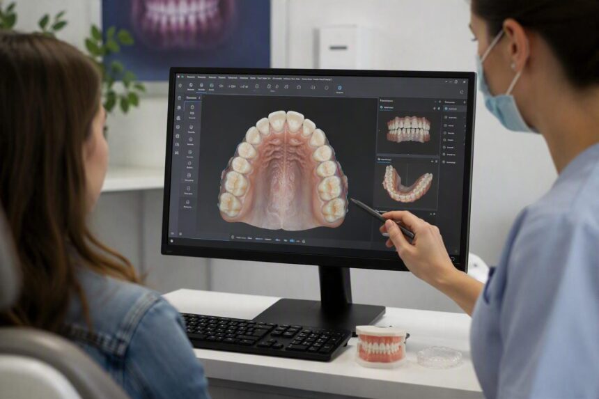
At the end of the wand is a small camera and a light source. When it goes over the teeth, it takes thousands of images each second and the software then joins these together to form a three-dimensional model which is assembled on the screen as you watch. If any areas are missed, they appear as gaps, and the dentist will simply go over those areas again until the model is complete.
It takes about a few minutes to scan both arches, after which there is a brief bite registration during which you have to close your teeth together so that the software can record how the upper and lower teeth meet. Generally, patients say that the most difficult thing about it is keeping the tongue out of the way, though children find it easy, mainly since nothing tastes bad and the wand can be removed at any time if you need a break.
After it has been completed, the model can be rotated, enlarged and seen from angles which a mirror cannot provide, including from behind the teeth and from directly underneath.
Why Putty Impressions Faded Out
Traditional impressions proved effective for planning braces, and in some clinics they are still used for certain purposes. They also had habits which annoyed all concerned. The material would tend to drag a little when being removed and would distort the shape. Bubbles damaged the trays so that they had to be retaken. Gagging made the procedure difficult for sensitive patients and almost impossible for some children. The plaster models made from those impressions ended up filling up the shelves and became chipped after a number of years.
Digital files eliminate all of that. Nothing is distorted, nothing is broken, and a copy reaches the laboratory within minutes instead of having to travel across the city in a box.
Turning the Scan Into a Treatment Plan
The model acts as a working surface. With the help of planning software, the orthodontist is able to isolate each tooth and move it separately, so that its present position and the position it is intended to end up in are displayed side by side on the screen. Crowding is measured rather than guessed at. The amount of space needed for a rotated tooth is calculated in comparison with the space that is available, and this influences the decisions regarding expansion, reshaping the contact points or, in a number of cases, the removal of a tooth.
Movement also has to be carried out in sequence, since all the teeth can’t move at the same time; the treatment plan therefore specifies which teeth move first, which will act as anchors, and where the bite needs to be corrected together with the alignment. Many patients are shown a simulation at this stage, in the form of a short animation that plays from their present position to the position to which the plan aims for them to reach.
The animation is worth a fair comment since it presents a plan rather than a promise. Because biology is different from person to person and teeth react at different speeds, the treatment is checked and modified at each appointment.
Planning Metal Braces From a Digital Model
The position of the bracket has a great effect on fixed braces. If a bracket is bonded a fraction of a millimetre away from where it should be, this alters the angle to which the tooth will eventually settle, and small discrepancies along the entire arch result in additional finishing work being required later.
With digital planning, brackets can be positioned on the virtual model first, the software then showing how the archwire will move through them. Occasionally, clinics transfer these positions into the mouth by using a custom tray produced from the same file; this method is called indirect bonding. Whether it is metal braces or clear braces, the ceramic brackets benefit since they operate on the same mechanical principles as the steel ones.
Planning Invisible Braces From the Same File
The aligner treatment places even more reliance on the scan since the trays are made directly from it. The anticipated movements are broken down into small steps, each of these steps corresponding to one tray. Wearing the trays in turn moves the teeth through the various stages of the treatment.
Attachments are also provided in this case. Small, tooth-coloured bumps are bonded onto certain teeth so that the aligner has something to grasp onto, enabling it to rotate or push a tooth rather than just slide over it. The shape and position of these bumps are determined on the model well before the first tray is worn.
A person considering invisible teeth braces as against fixed braces can get a useful advantage by looking at both options on the same scan, with just one visit and one medical record showing the two methods. This makes it much easier to talk about matters of discretion, the need for daily discipline and the complexity of the case than anything in a leaflet.
What Sits Alongside the Scan
Scanners are able to record the surface only, not the roots or the bone. To arrive at a complete assessment, orthodontists combine the scanner model with a clinical examination, photographs, and radiographs. In cases where this is necessary, for example, with impacted teeth or a complex relationship between the jaw, CBCT imaging is used, as it provides a three-dimensional view of the position of both the bone and the roots. Imaging is only prescribed when clinically required and in accordance with standard radiation protection procedures.
Assessment of gum health takes place before any procedure begins. It is not advisable to move the teeth through inflamed tissue, so scaling and a period of better at-home care are often carried out first.
Checking Progress Without Starting Over
Scans can be carried out during treatment and superimposed onto the original ones, thus showing how far each tooth has actually moved. If progress is found to be lagging behind the planned amount, adjustments are made earlier rather than at the next full review.
Files of stored items also retain their value at the end. Retainers are made from a scan taken when the braces are removed, and if a retainer is lost or cracked, a replacement can usually be produced from the file on record.
What This Changes for the Patient
The appointments become shorter and a great deal more comfortable since patients can look at their own teeth on a screen rather than trying to imagine what the dentist is saying, a method which in turn makes it easier to reach a decision. As a result, there being fewer remakes leads to fewer visits being fitted into working weeks.
The situation has also improved. The impressions are sent to the laboratory on the same day that they are taken, and the appliances made from them are ready to use rather than having to be adjusted at the chair. It is no longer normal to have to wait weeks between taking an impression and fitting it.
In Dubai, portability is particularly important since people travel around, move house and change clinics, and it is much easier for a digital record to be passed on to a dentist who is treating them elsewhere than it is for a physical set of plaster models. At the Magnum Dental Clinic, intraoral scanning is used as the first step in carrying out an orthodontic assessment and the same file is then used for planning, manufacturing and for retention.
Conclusion
Digital scanning has not altered the biology of tooth movement; what has changed is the quality of information that the orthodontist works with and the clarity with which that information can be communicated to the patient who is in the chair. Braces still take several months, still require regular adjustments, and still depend on patients fulfilling their part of the agreement. Starting with an accurate three-dimensional model simply eliminates a degree of guesswork, regardless of the route that treatment follows. Ask to see your scan during the consultation, since viewing your teeth from angles that you have never seen before makes it much easier to understand the entire treatment plan.
Contact Us
Orthodontic consultations are held at four locations in Dubai every day of the week by Magnum Dental Clinic.
- Dubai Silicon Oasis, SIT Tower: +971-54-336-3340
- Jumeirah Village Circle, Arcadia Tower: +971-50-719-3374
- Arjan, Al Barsha South: +971-55-890-8653
- Jumeirah Lake Towers, Indigo Tower: +971-54-452-2332