When a tooth must be removed, many people hope to replace it with a dental implant as soon as possible. However, the timing isn’t the same for everyone. Dentists weigh details you can feel and some you cannot see, from the shape of the tooth socket and the thickness of the facial bone to bite forces and gum biotype. Imaging such as a CBCT scan, the quality of the remaining alveolar ridge, and whether infection is present all guide the plan. Understanding the difference between placing an implant at the time of extraction and waiting through a healing phase helps you talk with your dentist confidently about bone grafts, temporary tooth choices, and what protects successful osseointegration over time.
-
Immediate vs Delayed Implant Placement After Extraction
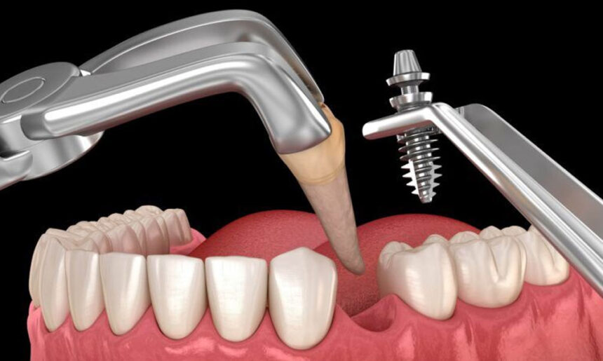
Immediate placement means placing the titanium implant fixture in the socket at the same visit as the extraction. Early or delayed placement schedules the implant after the site has partially or fully healed. Immediate placement can shorten the timeline and help preserve soft tissue contours, especially in the front of the mouth. It still requires accurate three-dimensional positioning for the future abutment and crown, along with strong primary stability in the bone. Waiting allows infection to resolve, supports guided bone regeneration when needed, and can increase predictability in areas with thin or damaged socket walls or limited bone volume.
Residents who want a deeper local overview can look into dental implants in Franklin to see how area clinicians stage imaging, grafting, and temporization. You will read about why a surgical guide may be used for more precise implant placement, how a healing cap or provisional crown can shape the gumline, and why some molars are better served by delayed placement due to heavier bite loading. This context will help you frame questions about your own tooth position, bone density, and timeline expectations.
-
Conditions That Favor Immediate Placement
Immediate placement tends to work well when the socket walls are intact, any existing infection has been appropriately managed, and CBCT imaging shows adequate bone to anchor the implant beyond the tip of the old root. A thicker facial bone plate and sufficient keratinized tissue support esthetic stability around the future crown margin. Primary stability needs to be achieved at surgery to keep micromotion low while bone grows onto the implant surface. In many cases involving front teeth after trauma, a dentist can remove a tooth that cannot be restored, place the implant slightly toward the palate for ideal alignment, add particulate bone graft to fill small gaps, and place a healing abutment or a carefully adjusted provisional that avoids biting forces.
-
When Waiting Yields a More Predictable Implant
Extraction sites with significant infection, a missing facial bone plate, or a thin tissue biotype often benefit from staged treatment. Your dentist may first perform socket preservation using a bone graft and a collagen membrane to maintain ridge width. In the upper back jaw where the sinus floor sits close to the roots, a sinus lift may be planned before or during implant placement to regain vertical bone height. Heavy bruxism, uncontrolled diabetes, and smoking increase the risk of early complications, so a delayed approach that stabilizes gum health and reduces inflammation can be safer. These steps allow the site to accept the implant in stronger bone with a more suitable shape and healthier soft tissue.
Consider a typical scenario: a cracked upper molar with a draining abscess and very thin bone between the root and the sinus. The tooth is removed as gently as possible, the socket is cleaned, and a graft is placed to preserve the ridge and prevent collapse. Months later, after healing and a follow-up CBCT scan, the dentist performs a crestal sinus lift to add a few millimeters of bone, then places the implant with good stability. The tradeoff is time, but the gain is a more predictable foundation for the abutment and crown in a chewing area that experiences heavy forces.
-
Temporary Tooth Choices During Healing
During healing, you still need a way to fill the space. Options include an Essix retainer with a tooth, a lightweight flipper partial, or a bonded Maryland bridge for some front teeth. In immediate cases with excellent stability and no heavy bite in the area, a provisional crown that does not bear biting forces may be attached to help shape the gumline. The tradeoff is clear: a removable temporary is simpler and keeps pressure away from the implant but may feel bulky; a fixed provisional looks and feels more natural but must be meticulously adjusted to avoid any chewing contact that could disrupt osseointegration.
For example, if a central incisor fractures and the bone and gum are healthy, an immediate implant with a carefully adjusted provisional can preserve the scalloped papillae and guide soft tissue healing. If the site lacks support or the bite is deep, a dentist may instead use an Essix retainer with an ovate pontic that lightly molds the tissue without loading the implant. Whichever route is chosen, meticulous hygiene is crucial. Remove and clean removable appliances daily, brush gently around healing caps, and avoid sticky foods that could dislodge a temporary or irritate sutures.
Aftercare That Protects Osseointegration
The first days focus on protecting the clot and minimizing movement. Cold compresses, a soft-food diet, and avoiding straws and tobacco support healing. Follow your dentist’s directions on rinsing and medications, and keep a soft toothbrush away from sutures until you are cleared. As the site stabilizes, gently clean around the healing abutment and adjacent teeth. After the final crown is placed, use interdental brushes or a water flosser to clean the junction between the crown and abutment, and schedule regular maintenance visits. Your dentist will check bite contacts, monitor crestal bone with periodic radiographs, and look for early signs of peri-implant mucositis, such as bleeding or swelling. For patients who clench or grind, a nightguard can reduce overload on the implant-supported crown.
In short, the right timing for an implant after extraction depends on the specific site, your systemic health, and how your bite loads the area. Immediate placement can preserve contours and shorten the calendar in ideal sockets, while a staged plan with grafting or a sinus lift can build a sturdier foundation when bone or soft tissue is compromised. Ask about your CBCT findings, whether socket preservation is recommended, and how a temporary tooth will be handled. A clear plan with carefully organized steps protects osseointegration and sets up the abutment and crown for lasting function and esthetics.